Prostate MRI
What is MRI of the Prostate?
Magnetic resonance imaging (MRI) is a noninvasive medical test that physicians use to diagnose and treat medical conditions.
MRI uses a powerful magnetic field, radio frequency pulses and a computer to produce detailed pictures of organs, soft tissues, bone and virtually all other internal body structures. MRI does not use ionizing radiation (x-rays).
Detailed MR images allow physicians to evaluate various parts of the body and determine the presence of certain diseases. The images can then be examined on a computer monitor, transmitted electronically, printed or copied to a CD.
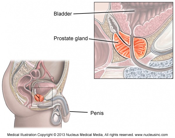
The prostate gland is part of the male reproductive system. It is located in front of the rectum and below the bladder, where urine is stored, and surrounds the first part of the urethra, the tube that connects the bladder with the tip of the penis and carries urine and other fluids out of the body. The prostate helps make the milky fluid called semen that carries sperm out of the body when a man ejaculates. Ultrasound and MRI are the most commonly used techniques to image the prostate gland.

What are common uses of a Prostate MRI?
The primary indication for MRI of the prostate is the evaluation of prostate cancer. The test is commonly used to evaluate the extent of prostate cancer in order to determine if the cancer is confined to the prostate, or if it has spread outside of the prostate gland.
Occasionally, MRI of the prostate is used to evaluate other prostate problems, including:
- infection (prostatitis) or prostate abscess.
- an enlarged prostate, called benign prostatic hyperplasia (BPH).
- congenital abnormalities.
- complications after pelvic surgery.
What happens during a prostate MRI?
You will be asked to change into a hospital gown and lie on a table attached to the MRI machine. The technician will place a specific coil around your pelvis to produce images. A small needle will be inserted into a vein in your arm or hand; this will be used to give the following medications:
- Buscopan, which helps to reduce the movement of the bowel. Bowel movement during the scan can affect the quality of the images of the prostate gland. It is important that the images are as clear as possible.
- Gadolinium contrast medium, sometimes just called ‘contrast’, which can help show any cancer in the prostate gland. Not every radiology practice will use contrast medium.
- Sedatives, to make you feel sleepy and relaxed, if you are one of the 10% of people who become claustrophobic in the MRI machine.
How Long Is the Prostate MRI Exam?
In most cases, the procedure takes 45 to 60 minutes, during which several dozen images may be taken.
What are the benefits of a Prostate MRI?
- MRI is a non-invasive imaging technique that does not involve exposure to ionizing radiation.
- MR images of the soft-tissue structures of the body including the prostate and other pelvic structures are clearer and more detailed than with other imaging methods. This detail makes MRI a valuable tool in early diagnosis and evaluation of the extent of tumors, such as prostate cancer.
- MRI has proven valuable in diagnosing a broad range of conditions, including cancer, conditions such as benign prostatic hyperplasia and infection.
- MR spectroscopy can examine the chemical makeup of the prostate which can be helpful in identifying prostate cancer. MR diffusion and MR perfusion imaging can evaluate other tissue properties of normal prostate tissue and prostate cancer.
- MRI enables the discovery of abnormalities that might be obscured by bone with other imaging methods.
- The contrast material used in MRI exams is less likely to produce an allergic reaction than the iodine-based contrast materials used for conventional x-rays and CT scanning.
What are the Risks of a Prostate MRI?
- The MRI examination poses almost no risk to the average patient when appropriate safety guidelines are followed.
- If sedation is used, there are risks of excessive sedation. However, the technologist or nurse monitors your vital signs to minimize this risk
- Although the strong magnetic field is not harmful in itself, implanted medical devices that contain metal may malfunction or cause problems during an MRI exam.
- Nephrogenic systemic fibrosis is currently a recognized, but rare, complication of MRI believed to be caused by the injection of high doses of gadolinium-based contrast material in patients with very poor kidney function. Careful assessment of kidney function before considering a contrast injection minimizes the risk of this very rare complication.
- There is a very slight risk of an allergic reaction if contrast material is injected. Such reactions usually are mild and easily controlled by medication. If you experience allergic symptoms, a radiologist or other physician will be available for immediate assistance.
What are the limitations of MRI of the Prostate?
The presence of an implant or other metallic object sometimes makes it difficult to obtain clear images. Patient movement can have the same effect.
A very irregular heartbeat may affect the quality of images obtained using techniques that time the imaging based on the electrical activity of the heart, such as electrocardiography (EKG).
MRI cannot always distinguish between cancer tissue and inflammation or presence of blood products within the prostate, which sometimes occurs related to a prostate biopsy. To avoid confusing the latter with the former on imaging, prostate MRI may be performed six to eight weeks after prostate biopsy, if possible, to allow remnants of bleeding to resolve.
