
Ultrasound
A service covered by RAMQ (Medicare).
Ultrasound or ultrasonography is a medical imaging technique that uses high frequency sound waves and their echoes. The technique is similar to the echolocation used by bats, whales and dolphins, as well as SONAR used by submarines.
The main advantage of ultrasound is that certain structures can be observed without using radiation. Ultrasound allows the study of multiple organs of the abdomen, the pelvis, the neck, thyroid, glands, liver, spleen, pancreas, kidneys, bladder, genitals but also the vessels (arteries and veins), the ligaments and the heart.
During pregnancy, it makes it possible to study the vitality and the development of the fetus, to determine the sex of the fetus or to detect abnormalities.
What are the benefits of an ultrasound?
- Ultrasound is a non-invasive, painless imaging technique.
- Ultrasound is widely available, low cost and easy to use.
- Because it does not use radiation, the side effects of radiation are not an issue. So, ultrasound is the preferred imaging modality for monitoring pregnant women and their unborn children.
- Real-time images are generated by ultrasound, so it is a good tool for guiding invasive needle biopsies.
- Ultrasound can display the movement and actual function of the body's organs and blood vessels.
What are the risks and limitations of an ultrasound?
- There are no known harmful effects of standard ultrasound imaging.
- The main limitation of ultrasound imaging is that it does not reflect clearly from bone or air. Therefore, it does not allow the study of all the body’s anatomy (ex.: bone, lung).
How does an ultrasound work?
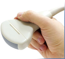
During the ultrasound examination, a machine called a transducer (probe) that both emits the sound and detects the returning echoes is placed on or over the body part being studied. The transducer (probe) sends the information it collects to the scanner, which generates the ultrasound image.
- It is a Technologist or a Radiologist who performs the exam.
- After registering you will be brought to a changing room where you will be told what items of clothing need to be removed for your examination and you will be asked to put on a patient gown.
- You will be given a key so you may lock your belongings in the changing room and bring the key with you.
- Once you are ready the technologist or assistant will bring you to the ultrasound examination room.
- A warm gel will be applied to your skin over the area that needs to be examined, to improve the contact with the probe.
- The Technologist will then glide it to the area of interest. The technologist will sometimes ask you to turn on your side, to take deep breaths, to stop breathing, etc...
- Sometimes, for a more specific study of certain areas (bladder, prostate, ovaries, and uterus) the probe will be introduced into the certain orifices (anus, vagina). For these exams you will be asked to sign a consent form.
- Do not hesitate to mention any problem or worries to the medical personnel during the examination.
- Depending on the area to be imaged, the examination lasts approximately 15 to 30 minutes. It is very quick.
- After the examination, you will be given a towel to remove the gel from your skin. You will be able to eat and drink normally.
- Results: The final report, prepared by the Radiologist will be addressed to your doctor within 48 to 72 hours. Your doctor will review the results with you and will recommend the appropriate treatment.
Ultrasounds offered at ViaMedica
Abdominal ultrasound
Thyroid ultrasound
Testicular ultrasound
Dating ultrasound
Ultrasound for children over 3 years of age
Pelvic ultrasound (+/- endovaginal)
Neck ultrasound
Obstetrical ultrasound
Surface (soft tissue) ultrasound
Doppler ultrasounds provide a more detailed study of the cardiovascular circulation.
Carotid doppler
Renal doppler
Hepatic doppler
Venous doppler of upper or lower limbs
Abdominal doppler

 For preparation instructions, click here